Cryo-electron microscopy (Cryo-EM) has revolutionized structural biology, providing unprecedented capabilities for determining the high-resolution three-dimensional structures of biological macromolecules, including proteins. This powerful technique allows researchers to visualize complex biological assemblies and challenging protein targets in near-native conditions, overcoming limitations of traditional methods like X-ray crystallography for certain sample types. Focusing on cryo electron microscopy for protein structure determination, this article explores how Cryo-EM enables detailed structural analysis and its significant impact on life sciences and drug discovery. To learn more about cutting-edge Cryo-EM services, please visit https://shuimubio.com/.
Understanding Cryo-EM for Protein Structure Determination
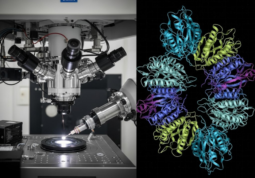
At its core, Cryo-EM involves flash-freezing biological samples in a thin layer of vitreous ice, preserving them in a near-native state without the need for crystallization that is often required for X-ray diffraction studies. An electron beam is then passed through the frozen sample, generating projection images. By collecting a large number of these two-dimensional images from many individual particles and using sophisticated computational algorithms, a high-resolution three-dimensional structure can be computationally reconstructed.
One of the primary applications of Cryo-EM for determining protein structures is Single Particle Analysis (SPA). SPA is a technique that specifically analyzes images of individual, identical particles (like purified protein molecules) to reconstruct their high-resolution 3D structure.
The general workflow for Cryo-EM SPA involves several steps: sample preparation (including protein expression and purification), negative staining characterization, grid preparation (freezing the sample), data collection using a Cryo-EM microscope, 2D particle picking from the images, 3D structure reconstruction, model refinement, and finally, data delivery. This comprehensive process allows for the visualization of proteins and other biological macromolecules at near-atomic resolution.
Advantages of Cryo-EM SPA for Proteins
Cryo-EM SPA offers several key advantages for protein structure determination:
· Preserves Samples in Near-Native State: Samples are flash-frozen in a hydrated state, which helps maintain their native conformation and biological activity.
· Captures Multiple Conformations: Unlike methods that require a single, stable crystal form, Cryo-EM can capture different functional states or conformations of a protein or complex present in the sample.
· Requires Less Sample: Cryo-EM often requires significantly less sample material compared to crystallography.
· Resolves Complex Structures: It is particularly well-suited for resolving the structures of challenging targets that are difficult to crystallize or are structurally heterogeneous, such as membrane proteins and large protein complexes.
· Determines Heterogeneous Protein Complex Structures: Cryo-EM can handle samples containing mixtures of related complexes, allowing for the resolution of distinct structures within the dataset.
These advantages make Cryo-EM an indispensable tool for obtaining detailed structural insights into a wide variety of proteins.
Diverse Protein Targets Resolved by Cryo-EM
Cryo-EM SPA is capable of resolving the three-dimensional structures of a broad range of biological macromolecules, including many types of proteins. Examples include:
· Proteins: This category encompasses various protein types.
o Membrane Proteins: This is a significant area where Cryo-EM excels due to the difficulty of crystallizing these targets. Examples include GPCRs (G protein-coupled receptors), ion channels, and transporter proteins. Specific examples mentioned include GPR75, TRPML1, and ASCT2.
o Enzymes: Cryo-EM can resolve enzyme structures, such as CYP51, C21, PolQ, and others.
o Ribosomes: The structure of ribosomes can also be determined using Cryo-EM.
· Protein-Nucleic Acid Complexes: Cryo-EM is effective for resolving the structures of complexes involving both proteins and nucleic acids. Examples include transcription complexes (like RNA polymerase bound to DNA) and viral capsid proteins complexed with viral RNA.
· Viral Particles: Cryo-EM is widely used to determine the structures of intact virus particles. This includes viruses like SARS-CoV-2, influenza virus, African Swine Fever Virus (ASFV), Human Herpesvirus 6B (HHV-6B), and rabies virus glycoprotein (VSV-GP). Resolving viral structures is crucial for understanding infection mechanisms and vaccine development.
This demonstrates the versatility of Cryo-EM in providing structural information for a wide array of proteinaceous entities and complexes.
Cryo-EM in Drug Discovery: Antibodies and Small Molecules
Cryo-EM for protein structure determination plays a critical role in modern drug discovery and development pipelines, particularly for antibody drugs and small molecule drugs.
Antibody Drug Development
In the development of antibody-based therapeutics, Cryo-EM offers several key applications:
· Antibody-Antigen Complex Structure Resolution: Cryo-EM can resolve the high-resolution 3D structures of antibodies bound to their target antigens. This provides crucial insights into how the antibody recognizes and interacts with its target, including the specific binding sites. Such structural information is invaluable for designing more effective antibody drugs. Case studies include the resolution of antigen-antibody complex structures.
· Mechanism of Action Studies: Cryo-EM helps in understanding how antibody drugs exert their effects by visualizing their interaction with targets and potential impact on downstream signaling pathways. This can reveal the molecular basis for antibody efficacy, such as how a neutralizing antibody blocks viral entry.
· Antibody Optimization and Design: Analyzing the structures of antibody-antigen complexes using Cryo-EM can reveal areas for optimizing antibody properties like affinity and specificity. It can also help in understanding conformational changes upon binding and identifying conformational epitopes, guiding antibody engineering efforts.
· Resolution of Membrane Protein and Complex Target Structures: Many antibody targets are membrane proteins (like GPCRs) or large, complex assemblies. Cryo-EM is highly effective at resolving the structures of these challenging targets, providing the necessary structural context for developing antibodies that bind to them effectively.
· Accelerating Drug Development: By providing detailed structural information quickly, Cryo-EM can significantly accelerate the antibody drug development process, allowing researchers to rapidly optimize antibody designs.
The ability of Cryo-EM to provide high-resolution structural insights into antibody-target interactions is crucial for the rational design and development of next-generation antibody therapeutics. To explore how Cryo-EM can advance your antibody drug research, please visit https://shuimubio.com/.
Small Molecule Drug Development
Cryo-EM is also making significant contributions to the development of small molecule drugs. Its applications in this area include:
· Target Structure Resolution: Cryo-EM can resolve the high-resolution structures of protein targets for small molecule drugs, such as membrane proteins and enzymes. For example, resolving the structure of GPCRs bound to small molecule ligands provides detailed insights into the binding site and interaction details, informing the design of highly selective and potent small molecules.
· Mechanism of Action Studies: Cryo-EM allows researchers to study the interaction mechanisms between small molecule drugs and their targets. Resolving the structure of a target protein bound to a small molecule agonist or antagonist can reveal how the drug activates or inhibits the target and affects downstream signaling. This structural information is vital for optimizing drug design and improving efficacy.
· Fragment-Based Drug Discovery (FBDD): Cryo-EM holds great potential in FBDD by revealing the specific interactions between small molecule fragments and protein targets. By determining the structures of protein-fragment complexes, researchers can quickly identify promising fragments and guide their optimization into lead compounds.
· Accelerating Drug Development: Similar to antibody development, the speed and high resolution of Cryo-EM structural analysis accelerate the small molecule drug discovery process, allowing for faster design and optimization cycles.
· Biased Ligand Studies: Cryo-EM is uniquely useful for studying biased ligands, particularly for GPCRs. Biased ligands selectively activate or inhibit specific downstream signaling pathways. Resolving the structures of GPCRs bound to biased ligands reveals their interaction mechanisms, aiding in the development of novel small molecules with more precise therapeutic effects.
· Resolution of Complex Targets: Cryo-EM's ability to resolve complex protein structures, such as membrane proteins and enzyme complexes, is invaluable for targeting these challenging proteins with small molecules.
By providing detailed structural information on targets and their interactions with small molecules, Cryo-EM provides powerful support for the rational design and optimization of small molecule drugs. Discover how Cryo-EM services can accelerate your small molecule drug discovery efforts at https://shuimubio.com/.
Complementary Cryo-EM Techniques for Protein Characterization
Beyond high-resolution SPA, other Cryo-EM techniques offer valuable insights into protein samples:
· Negative Staining (Negative Staining 2D): This technique provides a lower-resolution, but faster and more cost-effective, method for preliminary characterization of protein samples. Negative staining involves embedding the sample in a heavy metal salt stain, which provides contrast around the biological material. It is useful for assessing protein sample quality, including particle size, homogeneity, oligomeric state, morphology, particle density, flexibility, integrity, and conformational heterogeneity. It's often used as a screening step before proceeding to Cryo-EM SPA data collection. Negative staining is applicable for detecting homogeneity of AAV, exosomes, membrane proteins, viruses, and soluble proteins.
· MicroED (Microcrystal Electron Diffraction): While SPA focuses on individual particles, MicroED is used for resolving high-resolution structures from tiny protein crystals (microcrystals or nanocrystals) that may be too small for traditional X-ray diffraction. It can provide atomic resolution structures, and has been successfully applied to small molecule drugs, peptides, and proteins. Structures of proteins like Proteinase K have been resolved using MicroED. MicroED complements SPA by offering a route to atomic resolution for samples that form microcrystals, including proteins and peptides.
These techniques, alongside SPA, constitute a comprehensive suite of Cryo-EM services for studying protein structures and sample quality.
The Foundation: Protein Preparation and Analysis Services
Successful cryo electron microscopy for protein structure determination is highly dependent on the quality of the protein sample. High-quality, pure, and homogeneous protein is essential for obtaining high-resolution data. Recognizing this, leading Cryo-EM service providers often integrate comprehensive protein preparation and analysis services into their workflows.
These services typically cover:
· Protein Expression Systems: Utilizing various expression systems to produce the target protein, including E. coli, mammalian cells, insect cells, and cell-free systems, each chosen based on the protein's characteristics and requirements (e.g., size, post-translational modifications).
· Protein Purification: Employing multiple chromatographic techniques such as affinity chromatography, ion exchange chromatography, gel filtration (size exclusion chromatography), and RP-HPLC to isolate and purify the target protein to high homogeneity.
· Protein Quality Control (QC) and Characterization: Assessing the purity, integrity, and homogeneity of the purified protein using methods like SDS-PAGE, Western blot, mass spectrometry, thermal stability, and solubility tests. Protein-protein or protein-molecule binding analysis can also be performed.
· Protein Analysis Services: Offering specialized assays like SPR (Surface Plasmon Resonance), BLI (Biolayer Interferometry), and ELISA (Enzyme-Linked Immunosorbent Assay) to study protein interactions, kinetics, and concentration. These assays are critical for validating protein quality and functional activity before Cryo-EM.
Providing integrated protein services from gene sequence to purified, quality-controlled protein sample significantly enhances the chances of successful Cryo-EM structure determination.
Leading the Way in Cryo-EM for Protein Structures
Providers specializing in cryo electron microscopy for protein structure determination distinguish themselves through advanced technology, experienced teams, and proven track records.
A leading platform in Asia, established in 2017 from Tsinghua University, offers commercial Cryo-EM structure resolution services. Their core team comprises experts in life sciences, computation, IT, and the pharmaceutical industry. Key strengths highlighted include:
· Global Scale: Operating one of the largest commercial Cryo-EM platforms globally, equipped with multiple 300 KV Cryo-EM microscopes in Beijing and Hangzhou. The sources mention 8 microscopes currently equipped, with a potential for 12 in Beijing and 6 in Hangzhou for 300kV data collection.
· Extensive Experience: Possessing experience with over 400 Cryo-EM projects, having resolved over 150 protein structures. They have successfully resolved over 300 single-particle projects.
· High Resolution: Achieving excellent resolution, with a best resolution of 1.8 Å and reaching a breakthrough resolution of 1.4 Å. They have successfully elucidated structures of proteins as small as 51 kDa.
· AI-Driven Platform: Developing independent AI algorithms and software (like SMART and NanoSMART) to enhance the efficiency and accuracy of Cryo-EM data analysis and imaging. AI is used to accelerate data analysis and reduce machine time and required data volume.
· One-Stop Solution: Offering integrated services from protein expression and purification to Cryo-EM structure determination, aiming to streamline the process and reduce sample degradation issues during transport.
· Expert Team: Composed of PhD-level scientists from top institutions specializing in structural biology, protein science, and computational biology.
· High Project Success Rate: Successfully delivering over 80% of MicroED projects, achieving resolutions between 0.6-1.0 Å.
· Advanced Facilities: Utilizing state-of-the-art equipment including high-performance detectors, energy filters, spherical aberrations, and phase plates, with rigorous daily maintenance ensuring optimal performance and high uptime.
· Innovative Consumables: Developing proprietary consumables like Graphene grids (GraFuture™, GO, RGO) to address common challenges in sample preparation, such as preferred orientation and air-liquid interface damage, especially for small or low-concentration protein samples.
These factors collectively contribute to the capability to provide high-quality, efficient, and reliable services for protein structure determination using Cryo-EM. The platform offers 24/7 machine time services for data collection, with rapid response and dedicated technical support.
Successful projects and publications in top international journals, covering structures of ion channels, GPCRs, antigen-antibody complexes, spliceosomes, and other biological samples, validate the platform's capabilities. Specific examples of resolved structures include human GluN1-GluN2A NMDA receptors, histamine H1 receptor/Gq complex, and various SARS-CoV-2 related protein complexes.
Sample Requirements for Cryo-EM and MicroED
Obtaining high-resolution protein structures requires careful sample preparation. Specific requirements vary slightly depending on the Cryo-EM technique:
For Cryo-EM SPA:
· Protein Solution: Concentration typically ≥ 2 mg/mL, volume ≥ 100 µL, purity ≥ 90% (verified by SDS-PAGE, gel filtration). Minimize glycosylation or phosphorylation modifications if possible, and avoid repeated freeze-thaw cycles.
· Buffer: Minimize glycerol, salt ions, detergents, sucrose, and other organic solvents. Salt ion concentration ≤ 300 mM. Supply sufficient buffer volume (50-100 mL).
· Small Molecules (if co-complexed): Purity > 95%, required amount > 10 mg, soluble in DMSO or water to > 100 mM (or at least 1 mM if water solubility is poor). Affinity data (nanomolar level affinity to target protein) is recommended.
· Shipping: Ship fresh samples chilled or frozen samples on dry ice. Notify operations managers or sales colleagues at least 3 days in advance for fresh sample shipments.
For MicroED:
· Sample Type: Small molecules, peptides, or proteins.
· Sample Requirements: Crystalline form is required.
· Sample State: Powder, lumps, or other forms.
· Quantity: ≥ 5 mg is preferred, or a visible amount if not possible.
· Note: Samples must be stable crystals.
Detailed adherence to sample requirements significantly impacts the success and resolution of cryo electron microscopy for protein structure determination projects. For specific project inquiries and sample submission details, please contact https://shuimubio.com/.
Conclusion
Cryo-electron microscopy for protein structure determination has emerged as a transformative technology, enabling scientists to unveil the complex 3D structures of proteins and their assemblies at high resolution, often under near-physiological conditions. Through techniques like Single Particle Analysis, researchers can gain critical insights into protein function, mechanisms of action, and interactions with ligands, which are invaluable for fundamental biological research and applied fields like drug discovery.
Leading platforms, equipped with advanced Cryo-EM facilities, deep expertise, AI-driven technologies, and integrated protein science capabilities, are at the forefront of providing comprehensive solutions for high-resolution protein structure resolution. Their proven track record in resolving diverse and challenging protein structures demonstrates the power and potential of modern Cryo-EM.
As the demand for structural information grows in life sciences and pharmaceutical industries, Cryo-EM services continue to expand their capabilities, pushing the boundaries of resolution and tackling increasingly complex biological questions related to protein structures. Whether your research involves membrane proteins, large protein complexes, or protein-ligand interactions, Cryo-EM offers a powerful path to obtaining the detailed structural data you need.
To learn more about how Cryo-EM can support your research on protein structure determination and to access state-of-the-art Cryo-EM services, visit https://shuimubio.com/.