Determining the high-resolution three-dimensional structure of biological macromolecules is fundamental to understanding their function and developing targeted therapeutics. Cryo-Electron Microscopy (Cryo-EM), particularly Single Particle Analysis (SPA), has revolutionized structural biology by enabling the visualization of complex biological assemblies and previously intractable targets like membrane proteins in near-native states. However, a significant challenge within the field, especially for Cryo-EM SPA, remains the structure determination of cryo em small protein targets.
Proteins with low molecular weight pose unique hurdles for Cryo-EM SPA. The signal generated by smaller particles is weaker, leading to lower contrast in images and a poorer signal-to-noise ratio. This makes the crucial steps of particle picking and alignment more difficult. Furthermore, small proteins can be more susceptible to issues like background noise, strong preferred orientation on the grid, and damage at the air-water interface during sample preparation. Achieving a sufficiently high concentration of purified, homogeneous small protein sample can also be challenging. These factors often necessitate collecting exceptionally large datasets to compensate for the weak signal and limited orientations, increasing time and computational resources.
Despite these difficulties, advances in Cryo-EM technology, detector sensitivity, and image processing algorithms are increasingly making it possible to resolve the structures of cryo em small protein targets. Specialized techniques and services are vital to overcoming the intrinsic limitations associated with low molecular weight proteins.
Overcoming SPA Challenges for Cryo EM Small Proteins
One key area of innovation addressing challenges like preferred orientation, low concentration, and air-liquid interface damage, which disproportionately affect small proteins, is the development of advanced grid supports. For instance, graphene oxide (GO) and reduced graphene oxide (RGO) support films, marketed as GraFuture™, are designed to improve sample distribution, reduce orientation bias, and minimize damage, thereby enhancing the feasibility of resolving challenging samples, including those with low molecular weight or concentration. Using such specialized consumables can significantly improve data quality for cryo em small protein samples.
Furthermore, optimizing sample preparation protocols is paramount. For Cryo-EM SPA, requirements for protein samples typically include high purity (preferably >90%), sufficient concentration (often ≥2mg/mL), and a volume of at least 100ul. Buffer conditions are also critical, with recommendations to minimize organic solvents like glycerol and keep salt concentrations below 300mM. For small proteins, achieving and maintaining these concentration and purity levels can be particularly difficult, making expert sample preparation services invaluable. Careful handling to avoid freeze-thaw cycles and ensuring the sample is freshly prepared are also emphasized for successful grid freezing.
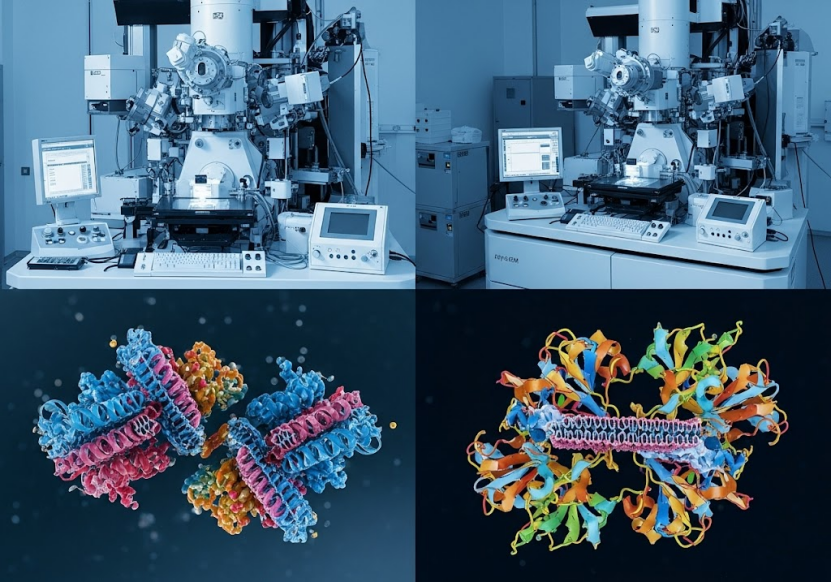
Specialized Cryo-EM facilities equipped with high-end 300kV electron microscopes featuring sensitive detectors, energy filters, and potentially spherical aberration correctors or phase plates, are essential for capturing high-quality data from weakly scattering samples like cryo em small protein particles. These advanced hardware components help to improve contrast and signal detection.
Beyond data collection, cutting-edge image processing software, particularly those leveraging AI technologies, plays a crucial role. AI-driven platforms can improve particle picking accuracy, facilitate better alignment, and enhance reconstruction quality even with noisy data from small particles. Such software can help to reduce the amount of data needed and increase overall efficiency.
Through the application of these advanced techniques and technologies, Cryo-EM SPA has demonstrated the capability to resolve structures of relatively small proteins. Notably, structures of proteins as small as 51kDa have been successfully elucidated, achieving impressive resolutions down to 1.8 Angstroms. This highlights the potential of modern Cryo-EM SPA to tackle samples at the lower end of the protein molecular weight spectrum traditionally considered challenging. The sources cite experience with over 400 Cryo-EM projects, resolving over 150 protein structures with a best resolution of 1.8 Angstroms, showcasing significant expertise in the field. They also mention successfully elucidating protein structures as small as 51kDa.
MicroED: A Complementary Approach for Very Small Structures
For even smaller samples, or when crystalline material is available, Microcrystal Electron Diffraction (MicroED) offers a powerful alternative. MicroED is particularly well-suited for determining the high-resolution structures of small molecules, peptides, and even small protein crystals. This technique utilizes electron diffraction from microscopic crystals, often obtained from samples that do not readily form large crystals suitable for traditional X-ray crystallography.
MicroED can provide atomic-resolution structural insights, sometimes reaching resolutions between 0.6 and 1.0 Angstroms, which is excellent for understanding the precise arrangement of atoms in a molecule. This technique is valuable for small molecule drug discovery and structural analysis of challenging peptides or proteins that yield only micro- or nano-crystals. Sample requirements for MicroED typically involve crystalline material, either as powder or small clumps, with a mass of at least 5mg, though visible quantities may suffice if 5mg is not possible. The sample must be stable crystals.
Integrating MicroED capabilities with Cryo-EM facilities allows for a comprehensive approach to structural biology, addressing a wider range of sample types and sizes, including those that fall under the umbrella of cryo em small protein or related small biological entities like peptides. The sources highlight the successful application of MicroED to protein structures (e.g., Proteinase K at 1.5 Angstroms), as well as peptides and small molecules, demonstrating its versatility for small sample structure determination. The development of specialized software like eTasED further enhances the application of MicroED on standard Cryo-EM systems.
Integrated Services for Challenging Samples
Successfully tackling the structure determination of cryo em small protein targets often requires more than just access to advanced microscopy. A fully integrated platform that covers the entire workflow, from gene sequence to purified, high-quality protein sample and finally to high-resolution structure, is essential.
Protein preparation is a critical bottleneck, especially for challenging targets like small proteins, membrane proteins, or those with low expression levels. Services offering diverse protein expression systems (E. coli, mammalian cells, insect cells, cell-free) and purification techniques (affinity chromatography, ion exchange, gel filtration, RP-HPLC) are vital for obtaining the necessary high-purity, concentrated sample required for Cryo-EM or MicroED. Expertise in handling difficult-to-express proteins, including membrane proteins (GPCRs, ion channels, transporters), is particularly valuable, as these can be small or require specialized handling. Rigorous quality control using methods like SDS-PAGE, Western blot, mass spectrometry, and thermal stability tests ensures the sample is suitable for structural studies.
Once the sample is ready, comprehensive structural services like "one-stop" SPA solutions and MicroED solutions guide projects through the entire process, from project evaluation and sample preparation to data collection, 2D particle picking, 3D reconstruction, model refinement, and final data delivery. Access to 24/7 data collection time on high-end microscopes further accelerates the process.
The combination of experienced scientists, advanced hardware, proprietary software, and an integrated workflow allows a platform to efficiently tackle challenging structural biology projects, including those involving cryo em small protein targets. Free project assessment and regular progress meetings ensure tailored solutions and transparency throughout the process.
Conclusion
Determining the structure of cryo em small protein targets presents unique challenges in structural biology, primarily due to the low signal-to-noise ratio and sample preparation difficulties inherent to low molecular weight samples. However, with advancements in Cryo-EM technology, specialized grid substrates, AI-driven image processing, and the availability of complementary techniques like MicroED for crystalline samples, these challenges are increasingly being overcome.
An integrated service platform that combines expert protein expression and purification, advanced Cryo-EM SPA capabilities, cutting-edge MicroED technology, and experienced scientists is crucial for successfully obtaining high-resolution structures of cryo em small protein and peptide targets. These platforms provide the necessary technical expertise and infrastructure to navigate the complexities associated with challenging samples, ultimately accelerating scientific discovery and drug development.
To learn more about how these cutting-edge technologies and comprehensive services can help you tackle the challenges of determining the structure of cryo em small protein and other challenging biological targets, please visit https://shuimubio.com/.