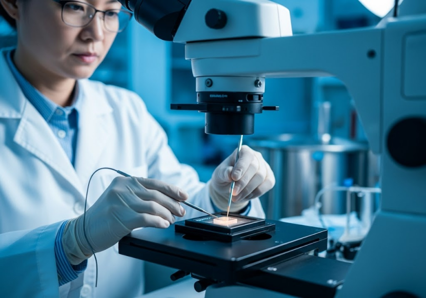
Solving Cryo-EM Challenges: Optimized Sample Preparation & Protocol Solutions

Cryo-electron microscopy (Cryo-EM) stands as a revolutionary technique in structural biology, enabling the visualization of biological molecules at near-atomic resolution. Unlike traditional methods like X-ray crystallography, Cryo-EM allows scientists to study macromolecules in a state closer to their native environment. This capability is crucial for understanding complex biological processes and developing new therapies. However, achieving high-quality Cryo-EM results often hinges on mastering two critical aspects: cryo em sample preparation and selecting the appropriate cryo em protocol.
Despite its power, Cryo-EM presents significant challenges. Obtaining high-resolution structures requires samples that are pure, homogenous, and correctly vitrified (frozen rapidly without forming ice crystals). Issues such as samples having a small molecular weight, low concentration, strong background noise, damage from the air-liquid interface, and preferred orientation can severely impede successful structure determination. Addressing these challenges requires specialized knowledge, advanced techniques, and optimized workflows.
The Critical Role of Cryo-EM Sample Preparation
Effective cryo em sample preparation is arguably the most crucial step in the Cryo-EM workflow. The quality of the sample directly impacts the ability to collect high-resolution data. Poor sample quality can lead to aggregated particles, low particle density on the grid, preferred orientation, or structural damage, all of which compromise the final structural model.
The goal of cryo em sample preparation for techniques like Single Particle Analysis (SPA) is to distribute individual particles evenly across a perforated grid, suspended in a thin layer of vitreous ice. This vitrification process is essential to preserve the native structure of the biological molecule. Standard preparation methods involve applying a small volume of sample onto a grid, blotting away excess liquid, and rapidly plunging the grid into liquid ethane or propane, which are cryogens (This is a standard practice in Cryo-EM not explicitly detailed in these sources, but necessary context for sample preparation discussion).
However, many biological samples are difficult to prepare effectively using standard methods. Membrane proteins, for instance, require detergents or nanodiscs to remain soluble, which can introduce high background noise or interfere with particle distribution. Small proteins or peptides may be difficult to visualize due to low contrast. Samples with high flexibility or multiple conformational states pose challenges for alignment and reconstruction. Viral particles or large complexes, while easier to see, require careful preparation to maintain their integrity and homogeneity.
The sources highlight that addressing these sample-specific challenges is paramount. For example, challenges like small molecular weight, low concentration, strong background noise, air-liquid interface damage, and preferred orientation need specific solutions.
Optimizing Cryo-EM Sample Preparation: Requirements and Solutions
Meeting the stringent requirements for cryo em sample preparation is vital for successful downstream analysis. The specific requirements vary depending on the sample type and the Cryo-EM technique being used.
For Cryo-EM SPA, general requirements for protein solution samples include:
· Concentration: ≥2 mg/mL.
· Volume: ≥100 µL.
· Purity: ≥90%, verified by methods like SDS-PAGE or gel filtration. Reducing glycosylation or phosphorylation may be beneficial, and repeated freeze-thaw cycles should be minimized, ideally using freshly prepared samples.
· Buffer: Should minimize glycerol, high salt ion concentrations (≤300mM), detergents, or sucrose. A sufficient volume (50-100mL) of buffer should also be provided for buffer exchange and concentration optimization.
For small molecule samples, requirements typically include:
· Purity: >95%.
· Quantity: >10 mg if possible.
· Solubility: Should dissolve to >100mM in DMSO or water; if water solubility is poor, at least 1mM is needed.
· Affinity data with the target protein (nanomolar level affinity recommended) is helpful. Samples should ideally be freshly prepared or sent on dry ice to avoid thawing.
Specific sample types also have tailored requirements for cryo em sample preparation. For Cryo-Characterization of nanoparticles like Lipid Nanoparticles (LNPs), liposomes, AAV, and other viral vectors:
· Liposomes: 1 mg/mL.
· AAV: Recommended E13 particles, at least 50 µL/sample.
· LNPs: Recommended concentration around 10 mg/mL; lower concentrations (e.g., 3 mg/mL) may not fill holes effectively. Sugar content should be <10% as high sugar affects contrast.
For Negative Staining & Negative Staining 2D:
· Protein Purity: >95% with no significant impurity bands or degradation bands on SDS-PAGE.
· Homogeneity: Should ideally show a single peak after gel filtration, with >90% homogeneity. Avoid re-concentrating after gel filtration to prevent aggregation.
· Volume & Concentration: 50-100 µL (single run) at 0.01-0.02 mg/mL.
· Buffer: No polysaccharides, DMSO, glycerol, or other organic substances. Salt ion concentration below 300 mM. Supply 15-20mL of buffer (50-100mL if gel filtration is needed) to help determine optimal sample concentration.
· Sample aliquots (15-20 µL/tube) are recommended to avoid repeated freeze-thaw cycles.
For MicroED, the sample must be a stable crystal:
· Sample Type: Small molecules, peptides, or proteins.
· Sample State: Powder, lumps, or other forms.
· Quantity: ≥5 mg; if not possible, a visually discernible amount.
To facilitate structure determination from gene sequence to high-precision 3D structure, an integrated "one-stop solution" is offered, including molecular cloning, protein expression, purification, and protein characterization. This platform aims to reduce the impact of sample transportation and overcome the difficulties in preparing hard-to-express proteins through standardized, automated engineering techniques. The protein preparation and analysis services include various expression systems such as E. coli, mammalian, insect, and cell-free systems, along with purification techniques like affinity chromatography, ion exchange chromatography, gel filtration, and RP-HPLC. Quality control methods like SDS-PAGE, Western blot, and mass spectrometry are also employed. This comprehensive approach addresses potential issues with sample quality before Cryo-EM grid preparation.
Furthermore, to tackle issues like preferred orientation and air-liquid interface damage, specialized materials like GraFuture™ grids (GO and RGO) have been developed. These graphene-supported membranes provide a potential solution for samples with small molecular weight, low concentration, high background noise, or those susceptible to interface damage and preferred orientation.
Diverse Cryo-EM Protocols and Services
The success of Cryo-EM also relies heavily on employing the correct cryo em protocol for data acquisition and processing. Different biological questions and sample types necessitate different Cryo-EM approaches.
1. Single Particle Analysis (SPA) Protocol SPA is a powerful cryo em protocol used to determine the high-resolution 3D structure of purified biological macromolecules like proteins, viruses, and protein complexes. The protocol involves collecting numerous 2D images of randomly oriented particles embedded in vitreous ice. Computer algorithms are then used to align and reconstruct these 2D images into a high-resolution 3D model. The workflow for SPA includes project consultation, evaluation, plan finalization, contract, protein expression/purification, negative staining characterization, sample vitrification & data collection, 2D particle picking, 3D structure reconstruction, model refinement, and data delivery. SPA is widely applied to study Protac, membrane proteins (GPCRs, ion channels, transporters), VLPs, peptides, and interactions between small molecules and target proteins.
2. Machine Time Service Protocol (24h) For researchers who have already prepared their samples (grids), 24-hour machine time service is available. This involves high-speed 300kV data collection using advanced electron microscopes like G3i, G4, and Totem. Currently, 8 x 300kV microscopes are available across platforms in Beijing (2) and Hangzhou (6). The service provides 24/7 response to booking requests, including expedited channels. The process includes online remote hole selection or selection guided by experienced scientists. Regular platform maintenance ensures optimal equipment status. The required sample is a frozen sample (grid) delivered at least one working day in advance, preferably transported in a liquid nitrogen tank.
3. MicroED Protocol MicroED (Micro-crystal Electron Diffraction) is a specialized cryo em protocol particularly useful for determining the high-resolution structure of small molecules, peptides, and proteins from microcrystals or nanocrystals. This technique requires crystalline samples. The workflow involves sample preparation (obtaining stable crystals), data collection from microcrystals, and structure analysis. An advantage is the capability to provide high-resolution structural insights from challenging small molecule and peptide samples. The proprietary eTasED software facilitates the application of MicroED technology on standard Cryo-EM systems without modifications, improving efficiency and accuracy. Success rates over 80% have been achieved with resolutions reaching 0.6-1.0Å.
4. Negative Staining & Negative Staining 2D Protocol Negative staining is an electron microscopy technique used to obtain low-resolution 2D projection images of macromolecules and complexes. The protocol involves staining the background with a heavy metal salt solution, making the unstained sample particles appear in contrast. This method provides preliminary information about particle size, homogeneity, oligomeric state, shape, particle density/sample concentration, protein structure, flexibility, sample integrity, conformation, and compositional heterogeneity at a lower cost than cryo-EM. Negative staining 2D specifically focuses on analyzing structures arranged on a 2D plane, common for viruses or protein complexes. It's often used as a preliminary assessment before proceeding to Cryo-EM SPA. Resolution typically ranges from 2-5 nanometers using transmission electron microscopy.
5. Cryo-Characterization Protocol Cryo-characterization utilizes cryogenic techniques to maintain the natural state of samples for high-resolution observation and analysis, particularly beneficial for proteins, liposomes, exosomes, and material interfaces. A specialized protocol is applied using the NanoSMART AI system, which automatically identifies nanoparticle features from images and generates detailed reports. This is particularly useful for characterizing LNPs, liposomes, AAV, and other viral vectors, providing data on size distribution, circularity, lamellarity, fill/empty ratio, and integrity. The AI can optimize identification even from lower-quality images.
Beyond Basic Protocols: Integrated Services for Complex Projects
Successfully navigating the complexities of drug discovery and life sciences often requires more than just a single Cryo-EM protocol. An integrated, "one-stop solution" approach can significantly streamline the process, from target identification to high-resolution structure.
This comprehensive approach includes protein preparation and analysis services, which are foundational for obtaining high-quality samples suitable for Cryo-EM or other structural methods. Beyond basic expression and purification, services extend to complex sample processing like complex incubation, molecular sieve chromatography for purity enhancement, and analysis of post-translational modifications like phosphorylation.
For targets amenable to crystallization, "one-stop" crystal structure analysis is available, covering the entire process from protein expression and purification to crystallization, data collection, and structure determination using X-ray crystallography. This is particularly useful for targets like KRAS or viral proteins.
In the field of antibody development, integrated services cover antibody discovery, including antigen preparation, library screening (phage display), antibody production, purification, and detailed characterization. Antibody characterization utilizes various protein assay technologies like SPR, BLI, and ELISA. These techniques measure binding affinity, kinetics, and protein concentration, providing crucial data that complements structural studies.
Driving Innovation with Technology and Expertise
Optimized cryo em sample preparation and advanced cryo em protocol execution are supported by cutting-edge technology and a highly skilled team. Founded in 2017, ShuimuBio emerged from Tsinghua University and became Asia's first commercial Cryo-EM structural analysis platform. The core team comprises leading life and computational scientists, IT experts, and pharmaceutical industry professionals.
The platform boasts a large-scale commercial Cryo-EM facility with 8 x 300 KV Cryo-EM microscopes. This extensive hardware infrastructure is designed for high-quality structural analysis and includes high-performance detectors, energy filters, spherical aberration correctors, and phase plates. Each microscope is meticulously maintained, ensuring high availability and operational efficiency.
Experience is key in navigating complex structural projects. The team has extensive experience with over 400 Cryo-EM projects, resolving more than 150 protein structures, reaching a best resolution of 1.8Å with SPA. Specific project examples include resolving structures as small as 51kDa and achieving a breakthrough resolution of 1.4Å. The MicroED service boasts an impressive 80%+ project success rate with resolutions down to 0.6-1.0Å.
Proprietary AI algorithms and software platforms significantly enhance the efficiency and accuracy of data analysis. The SMART software series leverages AI for Cryo-EM data analysis, reducing machine time and data volume. NanoSMART uses AI for automated identification and characterization of nanoparticles. eTasED applies MicroED technology seamlessly on standard Cryo-EM systems. These technological advancements, combined with deep expertise, address challenges inherent in both sample preparation verification and data processing, leading to faster, more reliable results.
Applications in Drug Discovery and Life Sciences
The ability to perform high-quality cryo em sample preparation and utilize diverse cryo em protocol options has profound implications across life sciences and drug discovery.
· Vaccine Development: Cryo-EM is invaluable in understanding virus structure and invasion mechanisms. It helps resolve structures of viral components (like SARS-CoV-2 S protein and ACE2 complexes) and can be used for vaccine quality control by assessing particle morphology, size, integrity, and aggregation. Studying antibody-vaccine antigen interactions helps optimize immunogenicity. Cryo-EM also aids in rapidly determining the structure of viral variants, informing vaccine design adjustments.
· Antibody Drug Development: Cryo-EM reveals high-resolution structures of antibody-antigen complexes, clarifying recognition mechanisms and binding sites. It's used to study antibody drug mechanisms of action, including how antibodies bind targets and influence signaling pathways. Structural insights from Cryo-EM inform antibody optimization and design. Importantly, Cryo-EM enables the resolution of complex membrane protein targets like GPCRs, which are frequent targets for antibody drugs.
· Small Molecule Drug Discovery: Cryo-EM provides high-resolution structures of drug targets, such as membrane proteins and enzymes, aiding in understanding drug action sites. It's used to study the interaction mechanisms between small molecules and targets, revealing how drugs activate or inhibit receptors and modulate pathways. Cryo-EM also shows potential in fragment-based drug discovery (FBDD) by detailing interactions between small molecule fragments and protein targets. It accelerates drug discovery by rapidly providing detailed structural information, crucial for optimizing drug design. The technology is also valuable for studying biased ligands and complex targets like GPCRs.
These application areas demonstrate the power of combining optimized sample preparation with flexible and advanced Cryo-EM protocols.
Conclusion
Mastering cryo em sample preparation and choosing the appropriate cryo em protocol are fundamental to successful Cryo-EM projects. The inherent challenges of biological samples require sophisticated solutions and expertise. By offering integrated protein preparation services, specialized materials like GraFuture™ grids, a range of Cryo-EM techniques (SPA, MicroED, Negative Staining, Cryo-Characterization, machine time), advanced AI platforms, and a team of experienced scientists, challenges related to sample quality, difficult targets, and complex protocols can be effectively overcome. This comprehensive approach supports cutting-edge research and accelerates structural studies across drug discovery and life sciences, leading to new insights and potential therapeutic advancements.
To explore how optimized cryo em protocol and expert cryo em sample preparation can benefit your research projects, from challenging membrane proteins to complex drug targets and viral structures, we invite you to learn more about these integrated solutions.
Discover advanced Cryo-EM services and overcome your structural challenges. Visit https://shuimubio.com/ for more information.