Understanding Cryo Electron Microscopy Principles with Shuimu BioSciences
In the realm of scientific visualization, Cryo Electron Microscopy (Cryo-EM) is the wizard that turns the invisible into the imaginable. At Shuimu BioSciences, we're not just practitioners of this magical art; we're its champions and educators, eager to share the principles that make Cryo-EM such a powerful tool in the arsenal of structural biology. Let's unfurl the scrolls and delve into the mystical world of Cryo-EM.
The Magic of Freezing Time
The first principle of Cryo-EM is the art of freezing time. Just as a photographer captures a fleeting moment, Cryo-EM immobilizes biological samples in a flash, freezing them in a thin layer of vitreous ice. This rapid freezing process, known as vitrification, halts molecular motion, preserving the sample's native state for detailed examination.
The Lens of the Atom
Cryo-EM uses a beam of electrons instead of light to visualize samples. Electrons have a much shorter wavelength than visible light, allowing for much higher resolution imaging – think of it as using a lens that can see individual atoms. This electron lens is the heart of the electron microscope, enabling us to see the intricate details of biological structures that were once beyond our reach.
The Beauty of 3D Reconstruction
With Cryo-EM, we don't just take a single snapshot; we collect a series of images from different angles. These images are then combined using sophisticated computational algorithms to create a three-dimensional reconstruction of the sample. It's like piecing together a puzzle, where each piece is a projection of the structure, and the final picture reveals the full, complex architecture.
The Power of 'Seeing' Macromolecules
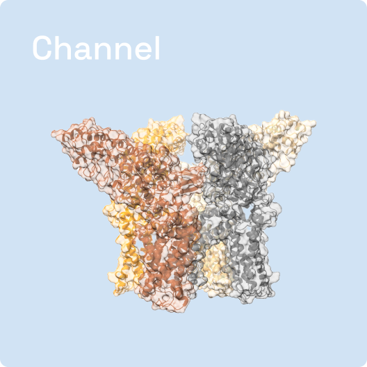
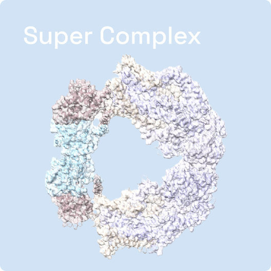
One of the most powerful aspects of Cryo-EM is its ability to visualize macromolecules, such as proteins and nucleic acids, at near-atomic resolution. This capability has revolutionized the field of structural biology, allowing scientists to 'see' these molecules in their natural state and understand how they function in the context of the cell.
The Precision of Single Particle Analysis
In single particle analysis, the sample is not a continuous sheet but thousands of individual particles. Each particle is imaged separately, and the best images are selected and aligned to reconstruct the three-dimensional structure of the particle. This method is the backbone of structural studies using Cryo-EM, providing unprecedented detail and precision.
The Art of Sample Preparation
The success of Cryo-EM relies heavily on the art of sample preparation. At Shuimu BioSciences, we've mastered the techniques to ensure that our samples are of the highest quality. From selecting the right buffers to optimizing the concentration of particles, every step is crucial in the journey to a successful Cryo-EM experiment.
The Future is in Focus
Cryo-EM is not just a tool for today's research; it's a beacon that lights the path to future discoveries. At Shuimu BioSciences, we're committed to advancing the practices and principles of Cryo-EM, ensuring that it continues to push the boundaries of what we can see and understand in the biological world.