Cryo-electron microscopy (cryo-EM) has revolutionized structural biology, allowing researchers to determine the three-dimensional structures of biological macromolecules and complexes at near-atomic resolution. A critical output of cryo-EM data processing, particularly from single particle analysis (SPA), is the cryo em density map. This map represents the distribution of electron density within the biological sample, providing the foundation for building accurate atomic models and gaining deep insights into biological function.
Understanding and effectively utilizing these cryo em density maps is paramount for downstream analysis, model building, and ultimately, advancing fields like drug discovery and vaccine development. Specialized software tools are essential for visualizing and interpreting these complex datasets.
The Power of Cryo-EM and Single Particle Analysis
Cryo-EM, particularly the Single Particle Analysis (SPA) method, allows for the determination of high-resolution three-dimensional structures of biological macromolecules such as proteins, viruses, and complexes. SPA works by taking numerous two-dimensional images of purified biological macromolecule particles preserved in a thin layer of vitrified ice, close to their native state. Computational algorithms then process and reconstruct these 2D images to generate a high-resolution 3D structure model. This process is crucial for studying proteins that are difficult to crystallize, including membrane proteins like GPCRs, ion channels, and transporters, as well as VLPs, peptides, and small molecules interacting with target proteins.
ShuimuBio, established in 2017, is a leading commercial cryo-EM structural determine service platform in Asia, with a core team comprising experts in life sciences, computational science, IT, and the pharmaceutical industry. They offer "one-stop" SPA solutions designed to tackle challenges such as small protein size, low concentration, high background noise, air-water interface damage, and preferred orientation. This comprehensive approach aims to deliver high-precision 3D structures starting from gene sequences. ShuimuBio's services cover the entire workflow, including protein expression and purification, negative staining characterization, sample freezing and data collection, 2D particle picking, 3D structure reconstruction, model refinement, and data delivery.
From Data Collection to the Cryo-EM Density Map
The process of obtaining a cryo em density map begins with high-quality data collection. ShuimuBio operates a large-scale commercial cryo-EM platform equipped with 300KV cryo-electron microscopes. They have 8 microscopes available, providing 24-hour data collection services in Beijing and Hangzhou. These state-of-the-art facilities, equipped with high-performance detectors, energy filters, spherical aberration correctors, and phase plates, are designed specifically for high-quality structural analysis and maintained regularly to ensure optimal performance.
Raw cryo-EM images captured during data collection contain projections of the biological particles from various orientations embedded in ice. These images are then subjected to extensive computational processing. This involves steps such as motion correction, CTF estimation, particle picking, 2D classification, 3D classification, and finally, 3D reconstruction. The outcome of the 3D reconstruction process is the cryo em density map. This map is essentially a volumetric representation of the electron density of the molecule or complex, reflecting the average structure derived from thousands or even millions of individual particle images.
The quality of the cryo em density map is directly related to the quality of the sample, data collection, and data processing. Factors like sample purity, homogeneity, and the resolution achieved during data collection significantly impact the final map. ShuimuBio emphasizes strict quality control throughout their process, particularly in protein preparation and characterization using techniques like SDS-PAGE, Western blot, mass spectrometry, thermal stability, and solubility tests to ensure samples are suitable for cryo-EM. They also utilize negative staining initially to check particle homogeneity, size, morphology, and concentration before moving to cryo-EM data collection.
ShuimuBio also addresses common challenges in sample preparation that can affect map quality, such as air-liquid interface effects and preferred orientation, by using innovative consumables like their proprietary GraFuture™ graphene support films. These films, including GraFuture™ GO (graphene oxide) and GraFuture™ RGO (reduced graphene oxide), help improve map quality, especially for small, low-concentration, or flexible proteins.
Furthermore, ShuimuBio leverages AI-driven platforms, including their self-developed SMART software series and NanoSMART system, to enhance the efficiency and accuracy of cryo-EM data analysis and processing. These AI tools can automate tasks like particle picking, reducing processing time and improving the quality of the generated density maps. The NanoSMART system is specifically designed for the characterization of nanoparticles like LNPs, liposomes, and AAVs, automatically identifying features and generating detailed reports from cryo-EM images.
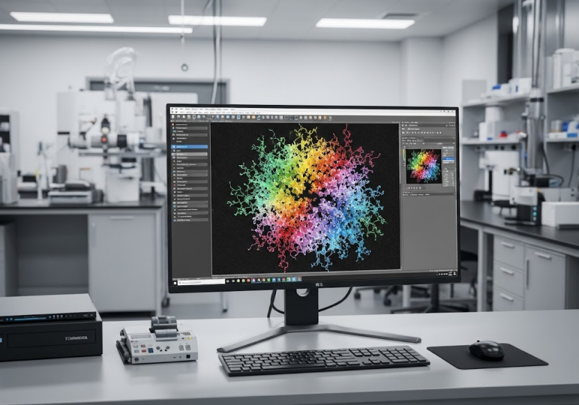
Visualizing and Interpreting the Cryo-EM Density Map: Introducing Chimera
Once a high-resolution cryo em density map is generated, the next crucial step is its visualization and interpretation. This involves displaying the volumetric density map in a 3D space and fitting an atomic model into it.
Software packages are indispensable for this task. One widely used program in the structural biology community for visualizing and analyzing cryo-EM data, including density maps, is Chimera, or its newer version, UCSF ChimeraX.
Chimera cryo em visualization tools allow researchers to:
· Load and display the volumetric cryo em density map at different contour levels. Adjusting the contour level is important for discerning structural features and ensuring appropriate fitting of the atomic model.
· Fit pre-existing atomic models (e.g., from the Protein Data Bank) or build new models directly into the density map. This is a key step in translating the electron density into an atomic representation of the biological molecule.
· Visualize and analyze specific regions of the map, such as active sites, ligand binding pockets, or protein-protein interfaces, providing crucial structural context for understanding function.
· Align and compare multiple density maps or atomic models to study conformational changes or differences between related structures.
· Generate images and animations for presentations and publications.
The interactive visualization provided by tools like Chimera cryo em is essential for validating the quality of the map, assessing the fit of the atomic model, identifying discrepancies, and ultimately refining the structural model to achieve the highest possible accuracy. A well-refined atomic model built into a high-resolution cryo em density map allows researchers to understand the precise arrangement of atoms and amino acid residues, which is critical for understanding biological mechanisms.
Applying Cryo-EM Density Maps in Key Research Areas
The ability to obtain and visualize high-resolution cryo em density maps has had a profound impact across various research and development fields. ShuimuBio's experience in over 400 cryo-EM projects, resulting in the determine of over 150 protein structures with resolutions reaching 1.8 Å, highlights the power of this approach. They have even achieved resolutions as high as 1.4 Å and successfully determined structures as small as 51kDa.
Here are some key application areas where visualizing cryo em density maps plays a crucial role:
· Vaccine Development: Cryo-EM enables the determine of virus structures at near-atomic resolution, which is vital for understanding infection mechanisms and designing vaccines. Visualizing the cryo em density maps of viral proteins, such as the spike protein of SARS-CoV-2 in complex with host cell receptors like ACE2, provides detailed structural information for vaccine design. ShuimuBio's work includes the determine of SARS-CoV-2 S protein structures and complexes. Cryo-EM also helps in vaccine quality control by visualizing particle morphology, size, integrity, and aggregation. Studies on antibody interactions with vaccine antigens, facilitated by cryo-EM density map analysis, help optimize immunogenicity.
· Antibody Drug Discovery: Determining the high-resolution structure of antibody-antigen complexes using cryo-EM density maps is essential for understanding how antibodies recognize and bind their targets. This structural information is critical for designing more effective antibody drugs. Visualizing the density map helps identify binding epitopes and interaction interfaces. Cryo-EM also supports the study of antibody drug mechanisms and the optimization and design of antibodies with improved affinity and specificity. ShuimuBio offers services for antibody discovery, including screening and characterization, which can benefit from cryo-EM structural analysis.
· Small Molecule Drug Discovery: Cryo-EM provides high-resolution structures of drug targets like membrane proteins (GPCRs, ion channels) and enzymes. Visualizing the cryo em density map of a target protein, often in complex with a small molecule ligand, allows researchers to see the precise binding site and interaction details. This structural information is invaluable for understanding the drug's mechanism of action, optimizing drug design for higher selectivity and potency, and supporting fragment-based drug discovery (FBDD). ShuimuBio has experience in determining the structures of various drug targets, including GPCRs and enzymes. Their "one-stop" solution from gene to structure is particularly beneficial for these projects.
Beyond Visualization: Structural Insights Lead to Breakthroughs
The ultimate goal of obtaining and visualizing a cryo em density map is to gain biological insights that can drive scientific discovery and drug development. By accurately modeling the atomic structure within the density map, researchers can investigate molecular interactions, conformational dynamics, and mechanisms of action at a level of detail previously unattainable for many challenging targets.
ShuimuBio's track record demonstrates the impact of their high-resolution cryo-EM services. Their work has contributed to numerous publications in top international journals, including structures of ion channels, GPCRs, antibody-antigen complexes, and spliceosomes. These publications highlight successful applications in understanding fundamental biological processes and identifying targets for therapeutic intervention.
Their repository of "shelf proteins," including various GPCRs, ion channels, transporters, and kinases, further showcases their expertise in producing high-quality samples suitable for cryo-EM structuraldetermine. Having access to well-prepared target proteins is a critical first step before generating a high-quality cryo em density map.
Conclusion
The cryo em density map is a cornerstone of cryo-EM structure determination, serving as the direct visual output of the experimental data and computational reconstruction. Its accurate visualization and interpretation using powerful software tools like Chimera (or ChimeraX), combined with expert knowledge, are essential for building atomic models and extracting meaningful biological insights.
These insights are driving innovation in diverse fields, from understanding the intricate structures of viruses for vaccine design to resolving complex membrane protein structures for targeted drug development.
For researchers and organizations seeking to unlock the power of high-resolution structural biology through cryo-EM and density map analysis, partnering with an experienced and well-equipped platform is crucial.
ShuimuBio offers state-of-the-art cryo-EM facilities, a team of top scientists, extensive project experience, and cutting-edge AI technology to provide comprehensive solutions for structure determination, from sample preparation to high-resolution density map generation and model building. Their commitment to achieving ultra-high resolutions and their focus on challenging targets make them a valuable partner in advancing structural biology research.
To learn more about how ShuimuBio's services can support your research projects and to explore the potential of high-resolution cryo-EM for your specific needs, please visit:
https://shuimubio.com/
Unlock the secrets held within the cryo em density map and accelerate your path to scientific discovery.